


CT Scan and MRI
CT Scan and MRI
This lesson aligns with NGSS PS4.C
Introduction
In the realm of modern medical diagnostics, two imaging technologies stand out as being highly effective for diagnosing and evaluating a wide range of health conditions: CT (Computed Tomography) scans and MRI (Magnetic Resonance Imaging). Both of these imaging techniques have revolutionized medical diagnostics, helping physicians visualize internal structures of the body in great detail. However, despite their common goal of providing images of internal body structures, CT scans and MRI work on entirely different principles, and they each offer unique advantages depending on the situation.This article explores both CT scans and MRI, explaining how each works, their benefits, and when each might be preferred for medical evaluation.
CT Scan (Computed Tomography)
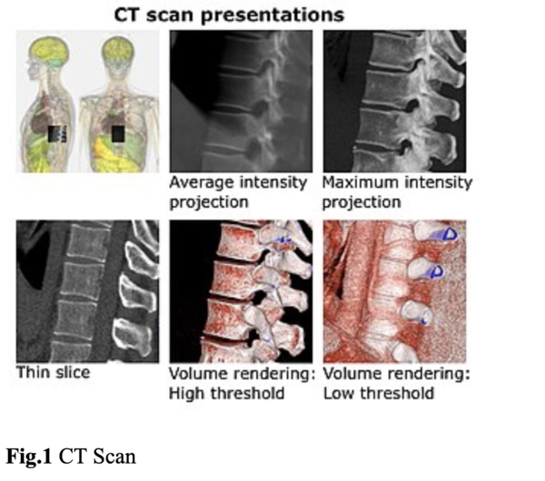
A CT scan, also known as computed tomography, uses X-rays to create detailed cross-sectional images of the body. This imaging technique allows doctors to see inside the body and examine bones, tissues, and organs from different angles.
CT scans are widely used in various medical fields, including emergency medicine, oncology (cancer diagnosis), and trauma care. They can quickly detect internal injuries, bleeding, tumors, or bone fractures and are often used to diagnose conditions such as cancer, heart disease, infections, and more.
How Does a CT Scan Work?
A CT scan works by taking multiple X-ray images from different angles around the body. These images are then processed by a computer to create cross-sectional "slices" of the body, which can be assembled into a three-dimensional representation of the internal structures.
Here’s a step-by-step breakdown of how a CT scan works:
- X-ray Tube Rotation: During a CT scan, the patient lies on a table that slowly moves through a large, circular machine called a CT scanner. Inside the scanner, an X-ray tube rotates around the patient, emitting X-rays at different angles.
- Detectors Capture Data: As the X-rays pass through the body, they are absorbed by different tissues in varying amounts. Dense structures like bones absorb more X-rays, while soft tissues like muscles and organs absorb fewer X-rays. Detectors positioned around the patient capture the X-rays that pass through the body.
- Computer Processing: The captured data is then sent to a computer, which processes the information and reconstructs cross-sectional images (slices) of the body. These slices can be viewed individually or stacked to create a detailed 3D image.
- Contrast Agents: In some cases, a contrast dye may be injected into the patient to highlight certain structures, such as blood vessels or organs, improving the visibility of specific areas.
MRI (Magnetic Resonance Imaging)
Magnetic Resonance Imaging (MRI) is a medical imaging technique that uses magnetic fields and radio waves to produce highly detailed images of the body's internal structures, particularly soft tissues.
MRI is particularly useful for imaging soft tissues like the brain, muscles, heart, and ligaments. It is commonly used to diagnose neurological conditions, joint and muscle injuries, heart problems, and diseases affecting soft tissues.How Does MRI Work?MRI works by exploiting the magnetic properties of hydrogen atoms, which are abundant in water and fat molecules in the body.
Here’s how an MRI scan works:
- Magnetic Field Creation: The MRI machine contains a powerful magnet that generates a strong, uniform magnetic field around the patient. When the patient is placed in the machine, the magnetic field causes the hydrogen atoms in their body to align along the direction of the field.
- Radiofrequency Pulses: Once the hydrogen atoms are aligned, the MRI machine emits short bursts of radiofrequency (RF) pulses. These pulses temporarily knock the hydrogen atoms out of alignment.
- Signal Detection: When the RF pulses stop, the hydrogen atoms return to their original alignment, releasing energy in the form of radio signals. The MRI machine’s detectors pick up these signals.
- Image Construction: A computer processes the signals to create detailed images of the body’s internal structures. The images can be viewed in cross-sectional slices, similar to CT scans, but with greater detail, especially for soft tissues.
Key Differences Between CT Scan and MRI
While both CT scans and MRI are essential tools in medical imaging, there are some key differences between the two techniques:
- Radiation: CT scans use X-rays, which expose patients to ionizing radiation. MRI, on the other hand, uses magnetic fields and radio waves.
- Imaging Focus: CT scans are excellent for imaging bones and detecting acute conditions like fractures and internal bleeding. MRI is superior for imaging soft tissues, such as the brain, muscles, and ligaments.
- Speed: A CT scan may take only a few minutes, while an MRI can take up to 30 minutes or longer, depending on the area being imaged.
- Cost: MRI scans are usually more expensive than CT scans due to the complexity of the equipment and the level of detail provided.
Conclusion
- A CT scan, also known as computed tomography, uses X-rays to create detailed cross-sectional images of the body.
- This imaging technique allows doctors to see inside the body and examine bones, tissues, and organs from different angles.
- Magnetic Resonance Imaging (MRI) is a medical imaging technique that uses magnetic fields and radio waves to produce highly detailed images of the body's internal structures, particularly soft tissues.
Related Worksheets:


Become a Help Teaching Pro subscriber to access premium lessons
Unlimited premium lessons Unlimited premium printables Unlimited online testing
- Already a Pro subscriber? Log in for access
- Browse free lessons
- Go back to previous page